Quantification of Bacteriophage by Spectrophotometry
Introduction
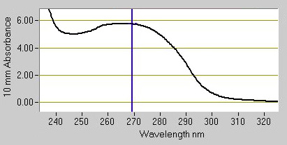
Measuring phage particles is a recurrent issue in phage display. The use of absorption spectrophotometry offers a rapid and simple method to measure the concentration of virion preparations. The technique was pioneered by George Smith and is based on the constant relationship between the length of viral DNA and the amount of the major coat protein VIII, which, together, are the major contributors of the absorption spectrum in the UV range. Here is a UV absorption spectrum of filamentous phage purified by PEG-precipitation and dissolved in TBS; the spectrum typically exhibits a shallow maximum around 269 nm:

The relationship between virion number and absorption is given by the following formula:

This formula was established by George Smith and can be found here. The calculation is based on the measurements of Day and Wiseman [Day, L.A. and Wiseman, R.L.: A comparison of DNA packaging in the virions of fd, Xf, and Pf1. In: Denhardt, D.T., Dressler, D. and Ray, D.S. (Eds.), The Single-Stranded DNA Phages. Cold Spring Harbor Laboratory, Cold Spring Harbor, NY, 1978, pp. 605–625]. At 320 nm phage chromophores have little absorption and this value is used to correct for light scattering from phage particles and non-phage particulate contaminants. Note as well the minimum absorption around 245 nm; when this minimum is absent from the spectrum, many contaminants are likely present in the preparation. This issue often happens when the preparation contains fewer virions and is an indicator that a second PEG-precipitation might be required.
Method
-
Blank the spectrophotometer between 240 nm and 320 nm with TBS 1x.
-
Replace the TBS with a dilution of the phage preparation in TBS 1x. As an example, if the phage was concentrated 10x times after the PEG-precipitation, dilute the preparation 10x time, back to the initial concentration in the bacterial culture.
-
Measure the absorption at 269 nm and at 320 nm and calculate the virion concentration with the above formula using the length of the phagemid vector + insert as the number of bases per virion.
Notes
-
If the absorption is above 2 O.D. at 269 nm, the preparation is too concentrated; use a higher dilution factor.
-
Undiluted preparations can be directly measured on a NanoDrop® spectrophotometer.
-
Virion preparations with A269 – A320 above 8.0 O.D. should be diluted for safe long term storage.
-
Always verify the quality of the spectrum; if the minimum around 245 nm is absent or the absorption at 320 nm is intense, the phage preparation is likely of low quality and the quantification erroneous.
NanoDrop is a registered trademark of Fisher Scientific.
Please send comments to info@abdesignlabs.com.
Copyright © 2013 Antibody Design Labs. All rights reserved. The reuse or reproduction of any of the information, design or layout contained in this web site without the permission of Antibody design Labs is prohibited.
